Assalamu Alaykum,
How you doing everybody?. Hope seemingly everything is rocking well and mesmerising. Well, would like to have a share with you regarding the most widely used radiographic assessment of root canal working length which is known as Ingles method in Endodontics
Without much ado, let's go to the main aspect of the write-up.
First of, as a clinician in dentistry dealing with Root Canal Treatment(RCT), it is better to the average length of each tooth's root as it helps in determining the working length during RCT.
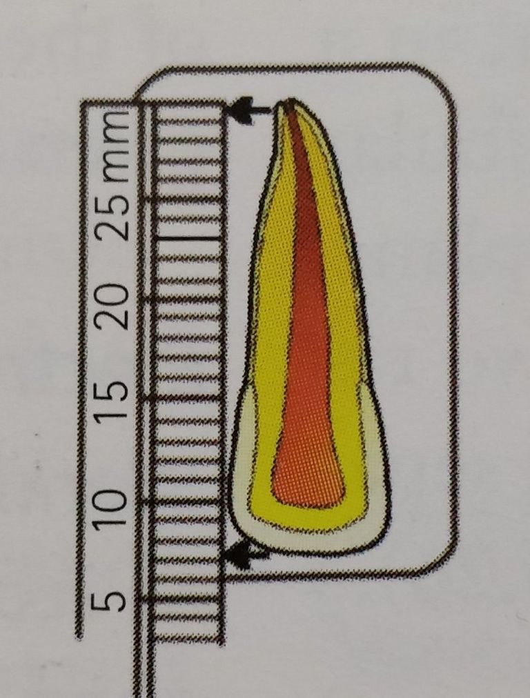
In INGLES' METHOD a diagnostic radiograph of the the tooth to be taken is the first thing to be done so as to know the radiograhic length and the radiograhic apex of the tooth in mm unit.
Secondly, subtracting 1mm from the radiograhic apex so as to know where to place your endodntic file and avoid exceeding the apical constriction level with the adjustment of the the silicon stopper accordingly on a stable coronal reference point.
After that a radiograph is retaken precisely a periapical radiograph. Three different scenarios may light like span out which can be as follows;
1- The file could be at exactly the apical constriction (also known as the minor diameter) which is the most small diameter of the root canal of a tooth. A file found in this anatomical location does not require any modifications as it is located at the required position both for "Cleaning and Obturation"
2- The file not reaching the apical constriction level then it will require the adding the missing length so as to reach the apical constriction known as the minor diameter with the appropriate adjustments to be made on the stopper of the file.
Take for instance if 2mm remain to reach the apical constriction then it would be added to reach out there.
3- The last but not least scenario is for a file to have exceeded the apical constriction level to the point of exiting the apical foramen which is the major diameter. This is corrected by substracting the exceeded length from the apical foramen up to the level of the apical constriction.
Take for instance, if from the radiograhic has exceeded 2mm from the apical foramen then 3mm should be subtracted from the exceeded file and adjusted on the silicon stopper respectively. The last remaining 1mm is from the radiograhic apex which is added to the extended part initially making 3 altogether.
Remember in both under- extension and over-extension with reference to the apical constriction, the stopper is always adjusted and also further radiograph is taken until the file is found on the appropriate location which is the apical constriction which is narrowest diameter portion of the root canal of every normal tooth.
For an accurate and more reliable working length determination a Ingle's radiograhic method combined with the use of electronic apex locator was reported to have more accuracy than relying on either of the two different methods separately.
That's all regarding the use of Ingle's radiograhic method in determination of root canal working length during RCT procedures.
Remain Blessed and Happy Blurting
Reference
Upvoted. Thank You for sending some of your rewards to @null. Read my last posts to make sure that BLURT burning is profitable for you. Before using this bot please make sure your account has at least 100 BP. Get more BLURT:
@ mariuszkarowski/how-to-get-automatic-upvote-from-my-accounts@ blurtbooster/blurt-booster-introduction-rules-and-guidelines-1699999662965@ nalexadre/blurt-nexus-creating-an-affiliate-account-1700008765859@ kryptodenno - win BLURT POWER delegation@ ctime/burn-bot-liquid-blurt