Hi everyone, on this post we would progress on mandibular fractures from where it was stopped in the previous post.
Mandibular fractures are quite interesting in learning due to the anatomical structures that are usually discontinued and broken into pieces right from the condyles to the symphysis end of the mandibular bone.
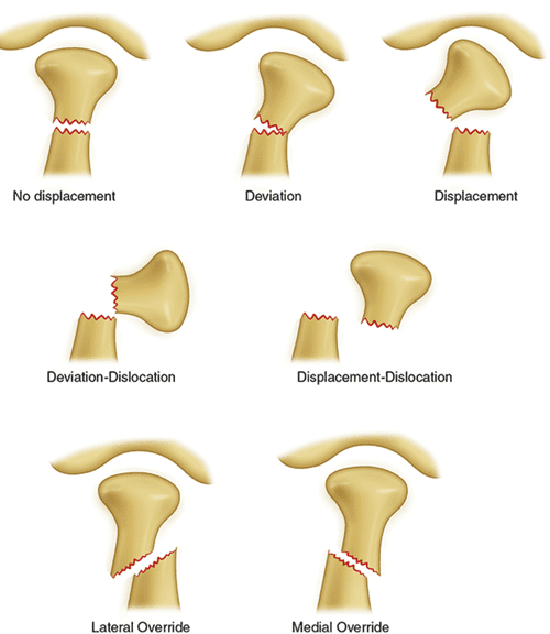
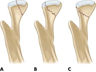
Condylar Fracture of the Mandible
This type of fracture of the mandible can involve a single condyle on one side (unilateral) or both sides (bilateral). Unilateral condylar fracture may cause restricted and an uncomfortable painful jaw movement. Swelling could be observed over the temporomandibular joint region and likewise harmmorrhage from the ear due to lacerations of the external auditory meatus. The hematoma may protrude downwards and backwards behind the ear, which may be mistook for Battle's sign (a sign of a base of skull fracture), though it is an uncommon case finding and for it to be spotted , intra-cranial injury must be excluded.
If the bones fracture fragments overlie each other this could results into the shortening of the height of the ramus and eventually results into gagging of the teeth on the fractured side (which there is premature teeth occlusion on the fractured side, and not on the non fractured side, giving rise to "open bite" that becomes increasingly obvious and worse to the unaffected side).
When the mouth is opened, there may be shifting of the mandible towards the fractured side. Two sided condylar fractures may cause the above signs and symptoms, but on both sides of the arch. Malocclusion and restricted jaw movement are mostly difficult and worse
Two sided mandibular body or parasymphysis fractures can be referred to as "flail mandible" at times, and can lead to involuntary posterior retrusion of the tongue which further obstructs the upper airway leading to eventual difficulty in breathing and swallowing on eating.
Punching and dislocation of the condylar bone during fracture upwardly across the roof of the glenoid fossa and into the cranial fossa is a very rare traumatic scenario.
"Bilateral condylar fractures combined with a symphyseal fracture is sometimes termed a guardsman's fracture. The name comes from this injury occurring in soldiers who faint on parade grounds and strike the floor with their chin"Source
Plain Film Radiography
In conventional radiology, plain films of the mandible would give clear information on diagnosis but had lower sensitivity and specificity together with lower reliable diagnostic information due to overlap of structures.
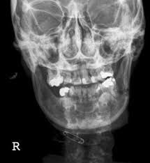
Views included anterior posterior (for parasymphsis), lateral oblique view and projection for (body, ramus, angle, coronoid process) and Towne's (condyle) views. Condylar fractures can be especially difficult to spot with confidence, partially due to the direction of condylar displacement or dislocation so multiple views of it are usually considered to be carried out during examination with atleast two views at perpendicular angulations.
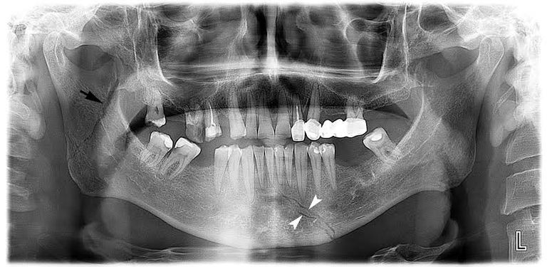
Panoramic Radiography
Panoramic radiographs are tomograms where the mandible is viewed wholly and a flat image of the mandible is depicted. In tomograms, mandibular structure appears in a 2-dimensional image, and as such fractures are easier to detect which makes it more reliable in accuracy and proximity to CT scans except in the condyle region of the mandible
Furthermore, broken, missing or malaligned teeth can often be easily spotted and appreciated on a panoramic image which is frequently lost and not available in plain films.
Medial/lateral displacement of the fracture segments majorly the condyle are difficult to ascertain on tomograms and as such the view is sometimes augmented and corroborated with plain film radiography or computed tomography to have more reliable diagnostic evidence and information regarding complex mandibular fractures.
Computed Tomography
Computed tomography has the most sensitivity and specificity of the imaging techniques and results because the facial bones can be viewed as layers through the skeletal framework and structure in any anatomical planes and views.
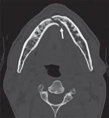
Images of the fractures can be reproduced into a 3-dimensional view, to give a better sense of the mobility, displacement and dislocations of various fragments. However, 3D reconstruction and viewing, can block and cover-up smaller fractures due to volume averaging and scaling, scatter artifact and surrounding structures that are just simply blocking and making the view of underlying areas unclear and not reliable enough for diagnostic assessment.
Consistent research has revealed that panoramic radiography is similar to computed tomography in its diagnostic accuracy for mandibular fractures and both are more superior in accuracy and reliability as compared to plain film radiograph.
The indications to use CT for mandibular fractures and dislocations differ by region, but it does not seem to make it more unique to diagnosis or treatment planning except for comminuted or avulsive type fractures, CT seems to have better clinician agreement on the location and absence of fractures when compared compared to panoramic radiography owing wide usage and acceptability for hard tissues imaging.
That's all as regards this post on mandibular fractures and their radiographical diagnosis in simple terms. On subsequent posts there would be discussions on classification of mandibular breakages with reference to anatomical locations and lots more. Thanks for the usual follow up and reading of the blogs.
Happy Blogging and Reading
Video from Dr Teeth YouTuber