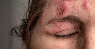
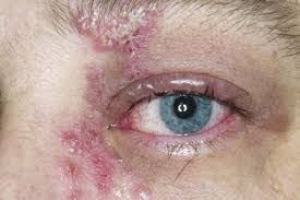
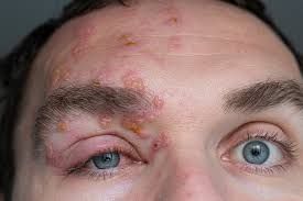
Unilateral rash appearance of the forehead, swelling of the eyelid, pain and reddish discolouration of the eyes.
Signs and Symptoms involving the skin of the face especially around the eye extending up to the nose tip, scalp and frontal face include the following clinically significant features and characteristics;
1- Viral prodrome
2- Preherpetic neuralgia
3- Rash, transitioning in stages from papules to vesicles to pustules and finally to scabs.
4- Hutchinson's sign: skin involvement of the tip of the nose, which indicates clearly nasociliary nerve involvement via the nerve supply. Though a positive Hutchinson's sign significantly indicates the likelihood of ocular complications as regards HZO, its absence does not exclude ophthalmic involvement after the reactivation of the virus in question.
5- Disseminated distribution in individuals with immunodeficiency
Herpes zoster ophthalmicus can also be observed during clinical examination during fluorescence staining through the use of cobalt blue light.
"Epithelial: punctate epithelial erosions and pseudodendrites: often have anterior stromal infiltrates. Onset 2 to 3 days after the onset of the rash, resolving within 2–3 weeks. Common."Source
"Nummular keratitis: have anterior stromal granular deposits. Occurs within 10 days of onset of rash. Uncommon
Necrotising interstitial keratitis: Characterised by stromal infiltrates, corneal thinning and possibly perforation. Occurs between 3 months and several years after the onset of rash. Rare."Source
Uveal
Anterior uveitis inflammation occurs in 40–50% of people with HZO within like 2 weeks of the onset of the rashes appearance.
Typical HZO keratitis and at least mild iritis is highly noticeable especially if Hutchinson's sign of the nose tip is positive which indicates the presence of vesicles upon the nose tip.
HZO uveitis is also associated with fatal complications such as iris decrease in size (atrophy) and secondary glaucoma from another disesase which are not uncommon.
Debilitating cataract may surface out at the late stages of this varicella zoster virus infection manifestation.
That's all as regards the post on symptoms and signs of Herpes Zoster Ophthalmicus infection of the eyes and facial skin. More would be better discussed on the anatomical supply of the branches of the trigeminal nerve which harbours the latent varicella zoster virus that is reactivated leading to the manifestation of Ophthalmicus Zoster infection in the facial region of the body tissues. Thanks for the usual support as it is.
Happy Blogging and Reading
Video from Dr Eye for you YouTuber


Remove the parasites... remove the problem...
CDS - The Truth about Chlorine Dioxide Solution
"The 'Universal Antidote' Documentary 'Chlorine Dioxide' The Miracle Mineral?"