Purpura
Bleeding into the skin or mucosa that results that results in violet or purple discolouration that varies based on its duration, does not blanch with pressure and later fade away over time.
Purpuric bleeding is divided into two groups;
1- Petechia
2- Ecchymosis
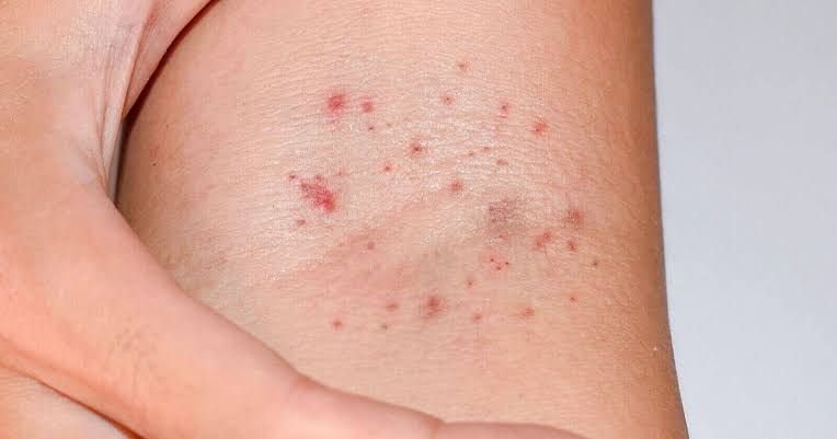
1- Petechia
Is defined as a tiny, 1mm to 2mm in diameter pinpoint non-blanchable macules which occurs due to rupture and damage to small blood vessels giving rise to red, purple or brown in colour and later fade away with time.
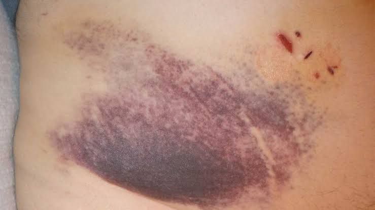
2- Ecchymosis
It is also a purpuric macule bleeding pattern which is greater than 3mm in diameter which occurs due to extravasated blood in the skin or mucosa and is non-blanchable. The colour may change from blue black to brown yellow or green before fading away.
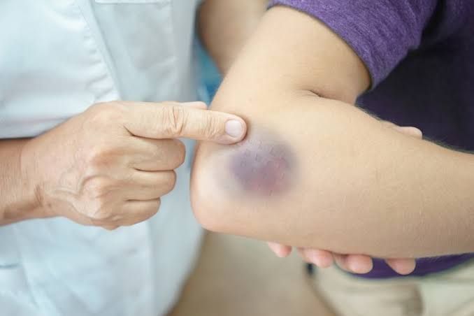
3- Hematoma
Hematoma refers to accumulation of extravasated blood relatively or completely within a specific space in the body structure.
The accumulated blood is usually completely or partially clotted and depending on lesion duration, it may undergo various degrees of organisation and colour patterns.
Hematoma following local anesthesia and dental extraction are most common complications in dentistry.
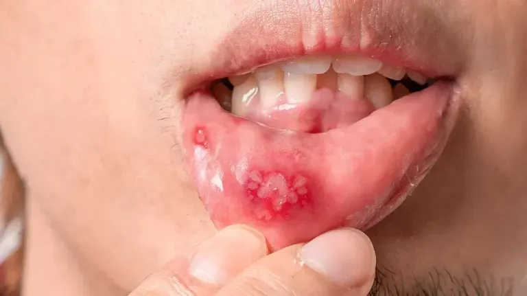
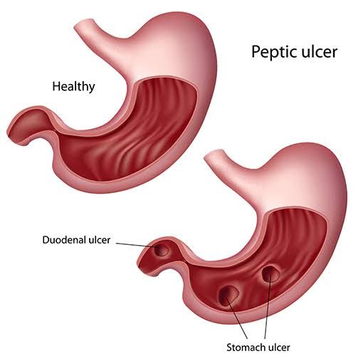
4- Ulcers
Ulcers are loss of continuity in the epithelium. The center of the lesion is initially red at the eruption of the lesion and later turns gray white after being covered with fibrin clot.
The surrounding border of the lesion may reddish in colour. Ulcers can be further classified according to their depth, border, shape, margin and tissue at its base.
Based on depth, ulcers can be superficial or deep. Superficial ulcers have less than 3mm dimension depth while deep ulcers have like 3mm and above depth.
Ulcers may be assymetric or symmetric forms according to the degree of the regularity of the borders and the margin can be smooth or crater-like when it is above the level of the normal mucosa.
Oral ulcers are the most common type of oral lesions such as canker sores, traumatic ulcers, drug-induced ulcers and so on.
Source
That's all as regards this post. In the subsequent posts, there would be more discussions on oral lesions. Thanks for the usual support and reading of the post.
Happy Blogging and Reading
Video from Medicosis Perfectionalis YouTuber