Most traumatic ulcers are etiology dependent, which means that once the causing agent has been removed or eliminated from further insults to the oral mucosa, the ulcer tend to or get resolved within 10-14 days provided no other factors were behind the appearance of the ulcer.
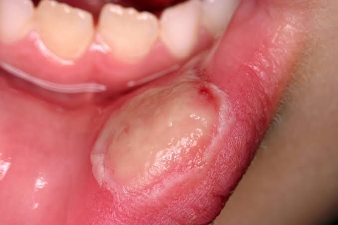
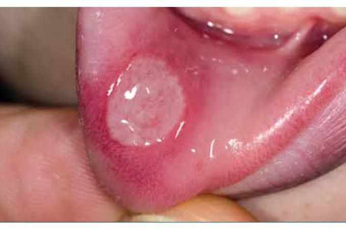
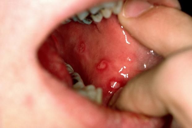
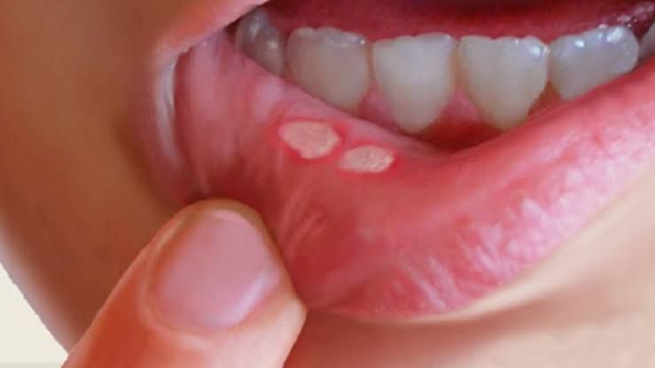
In most occurring cases the stimulus is identified and removed and the complain is mostly about pain and painful ulceration. The noticeable pattern of traumatic ulcers are either shallow or deep ulcers associated with border areas of erythema.
Due inflammatory response, an ulcer may appear to have a central yellowish or purulent exudate at its lumen. Sometimes the border of an ulcer maybe indurated depending on its duration.
Likewise a traumatic ulcer in the oral cavity maybe infected as a form of complication, if a prompt attention wasn't given.
Diagnosis of Traumatic Ulcers
Diagnosing traumatic ulcers mainly based on the history of trauma or insults which could be hot, cold, chemical or even ulcerations following the use of radiation therapy.
Mechanical trauma induced ulcerations usually follow a linear pattern of appearance. The depth of an trauma usually dependent on the nature of insult and its impact.
Generally the area surrounding and bordering the ulcers are often inflammed and in case of a repeated insults the traumatic factor are usually spotted around the vicinity of the ulcers such as a sharp-edged tooth, an ill-fitting dentures or over-filled tooth and so on
Differential Diagnosis of Traumatic Ulcers
Traumatic Ulcers can resemble any of the following anolamlies affecting the dental tissues;
(i) Carcinomatous ulcers e.g SCC ( Simple Squamous Cell Carcinoma Cancers)
(ii) Recurrent Apthous Stomatitis
(iii) Deep seated ulcers relating to opportunistic microbes
Histological Features of Traumatic Ulcers
Histopathologically a traumatic can appear under clinical examination as an area of surface ulceration which covered by fibrinopurulent exudates which comprises acute inflammatory cells, pus and intermixed with fibrin
Moreover, the stratified squamous epithelium may appear hyperplastic and wrinkled depicting areas of reactive squamous atypia.
Likewise, ulcer bed or base usually constitutes proliferation and formation of granulation tissues coupled with acute inflammatory and chronic inflammatory cells intermixed
Management of Traumatic Ulcers
1- Daily consumption of soft and balanced diet
2- Elimination of traumatic factor if available such as root stumps, malposed third molars, sharp cuspal edges of the teeth which can be filed and grounded.
Relining and rebasing of irritating dentures, restoration of fractured tooth and so on. If the etiological factor is actually removed then the ulcer will resolve and heal completely within 10-14 days.
3- Patient may be advised to rinse the hot saline for possible wear off of any availability
microbial pathogens.
4- Superficial application of antiseptic, analgesic and anaesthetic medication to remove symptomatic pain of the ulcer such as choline salicylate, Benzalkonium, and lignocaine hydrochloride.
This regimen is applied 10 minutes to food and 3-4 times a day.
5- For extremely large and deep ulcers which can be easily infected by food substrates and bacteria, penicillin could be a drug of choice for preventing infections, thereby assisting in faster healing.
6- Last but not least, patients suffering from self-inflicted trauma as an etiologic factor can be referred to a psychologist or psychiatrist after symptomatically managing the ulcer as the infliction can be due to mental or psychogenic reasons and in furtherance of diagnosis to rule out any mental health issues.
That's all as regards the significant clinical features of traumatic ulcers in oral medicine. More would be greatly discussed on other emerging ulcers on the oral cavity in the subsequent write-ups. Thanks for cool support and attention given to the post.
Happy Blogging and Reading 🦷🦷🦷🦷🦷
Video from Dr Ashank Mishra YouTuber


Telegram and Whatsapp
Telegram and Whatsapp